Kurskatalog forskarutbildning - VT24
-
Startsida
Ansökan kan ske mellan 2023-10-16 och 2023-11-15
Application closed
Skriv ut
Skriv ut
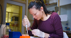
| Titel | Fluorescence microscopy: High content image acquisition and analysis |
|---|---|
| Kursnummer | 2973 |
| Program | Cellbiologi och genetik |
| Språk | Engelska |
| Antal högskolepoäng | 3.0 |
| Datum | 2019-09-09 -- 2019-09-20 | Kursansvarig institution | Institutionen för cell- och molekylärbiologi |
| Särskild behörighet | |
| Kursens syfte | Fluorescence microscopy is for most researchers an essential tool. Lately, this technique developed more and more from 'simply' acquiring good looking images towards complex, high content imaging techniques. High content imaging is defined by the large number of image data generated either from live cell imaging, or microscopy on fixed samples. Examples are z-stack imaging, cell migration, protein dynamics, multi-position imaging, tiling & stitching, whole mount imaging, and automated image acquisitions in screening assays. The purpose of this course is to make the participants familiar with all of these high content techniques; from acquisition to analysis and presentation. |
| Kursens lärandemål | After passing the complete course, the participants will be familiar with diverse high content fluorescence microscopy applications. They will be competent in designing and performing experiments involving high content fluorescence microscopy. The participants will be able to analyse their data using an image analysis software, and how to present the data in a scientific format. |
| Kursens innehåll | Basic principles of fluorescence microscopy. Confocal Laser Scanning Microscopy: z-stack, tiling & stitching. Live Cell Imaging: long term imaging, protein dynamics (FLIP/FRAP). Automated Image Acquisition Microscope: multi-well imaging, screening. Hands-on sessions with diverse high content imaging experiments. Image analysis: ImageJ/CellProfiler. Lectures and workshops. Poster presentation |
| Arbetsformer | The pedagogic learning activities in the course consist of lectures, research seminars, hands-on experience at the microsopes/imaging, group discussions, experimental design, data processing and poster presentation. |
| Obligatoriska moment | All activities (lectures, research seminars, microscope sessions, group discussions, data processing and poster presentations) are compulsory. If students are unable to attend they have to discuss with course organizer how this can be compensated. Compensation of microscopy sessions is not possible. |
| Examination | There will be 2 examinations. In the end of the first week, there will be an assessment of written versions of experimental designs involving high content imaging experiments and image analysis. In the second week, the participants will present a scientific poster showing the results of their high content microscopy experiments performed during that week. The poster presentations will be attended by course participants, lecturers, course assistants, and others that are interested. |
| Kurslitteratur och övriga läromedel | |
| Antal studenter | 12 - 18 |
| Urval av studenter | Selection will be based on 1) the relevance of the course syllabus for the applicant's doctoral project (according to written motivation), 2) date for registration as a doctoral student (priority given to earlier registration date) |
| Övrig information | The course takes place at Biomedicum, Solna Campus, room B0313. |
| Ytterligare kursledare | The course director is Florian Salomons, Department of Cell & Molecular Biology, Biomedicum Imaging Core (BIC). Email: Florian.Salomons@ki.se. Telephone 0852487395. |
| Senaste kursvärdering | Kursvärderingsrapport |
| Kursansvarig |
Florian Salomons Institutionen för cell- och molekylärbiologi 0852487395 Florian.Salomons@ki.se |
| Kontaktpersoner |
Matti Nikkola Institutionen för cell- och molekylärbiologi Matti.Nikkola@ki.se |