Kurskatalog forskarutbildning - VT24
-
Startsida
Ansökan kan ske mellan 2023-10-16 och 2023-11-15
Application closed
Skriv ut
Skriv ut
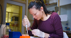
| Titel | Microscopy: improve your imaging skills |
|---|---|
| Kursnummer | 2871 |
| Program | Utveckling och regeneration (DevReg) |
| Språk | Engelska |
| Antal högskolepoäng | 4.5 |
| Datum | 2019-01-22 -- 2019-02-08 | Kursansvarig institution | Institutionen för biovetenskaper och näringslära |
| Särskild behörighet | The participants to this course must have used a microscope to acquire digital images of fluorescently labelled samples within the past 1 year. |
| Kursens syfte | The aim for this course is to improve the microscopy skills of students and researchers who have already used a microscope to acquire digital images of fluorescent samples and want to improve their skills. |
| Kursens lärandemål | At the end of the course, the participants will be able to: 1- Describe the difference between wide field and confocal microscopes as well as the different types of confocal microscopes 2- Pick the best combination of fluorophores for their experiment by matching their spectra with the microscope light source and filters, identify and eliminate bleed-through and cross-excitation problems 3- Explain objective specifications and limitations 4- Explain the theory behind settings the following parameters on a confocal or a wide field system to best match the requirements of their sample and reliably answer their scientific question: resolution, pixel size, averaging, scan speed, illumination power, detector gain and offset, camera readout rate, exposure time and camera binning 5- Explain which applications require a hardware or a software autofocus, a spectral detector, a resonance scanner, light sheet or two-photon microscopy or super resolution 6- Explain the advantages in using the automation of a microscope system to collect multidimensional data 7- Explain how to deal with images before publication in scientific journals 8- Run a simple image analysis on freeware (ImageJ/FIJI, Cell Profiler) and describe the imaging requirements for automated image analysis |
| Kursens innehåll | The students will learn the theory about the parameters and hardware used in in wide field and confocal imaging, how to identify and avoid imaging artefacts, deal with the challenges of imaging fluorescent volumes, as well as how to handle scientific images for publication. They will also learn about more advanced techniques. |
| Arbetsformer | Lectures, videos, peer review, image troubleshooting in groups. |
| Obligatoriska moment | Attendance to all sessions is compulsory. Any absence must be reported to the course leader in advance by e-mail. Absence from any part of the course (lectures, laboratory sessions, discussion sessions and exam) is generally not accepted but could in exceptional cases be compensated by a written additional assignment to ensure the learning outcomes of the day have been reached. If it is not possible to compensate, the student will be given a chance to complete the course by attending the missing sessions the following year. |
| Examination | The final mark (pass or fail) will depend on the results of: 1. The weekly assignments 2. The skills shown in each workshop 3. The written examination at the end of the course. The student has to show that all intended learning outcomes of the course have been reached. |
| Kurslitteratur och övriga läromedel | Handbook of Biological confocal microscopy, James Pawley Springer Editions 2006 |
| Antal studenter | 8 - 40 |
| Urval av studenter | The participants will be selected based on the usefulness of the course to their research project. The students must have been acquiring images on a microscope in the past 1 year. Their microscopy research project, as well as their previous microscopy experience must be described in writing in the application. |
| Övrig information | The detailed program can be found on the LCI website (https://ki.se/en/bionut/welcome-to-the-lci-facility) under Learning microscopy. Presence at the course is mandatory 3 days per week during 3 consecutive weeks, mostly (with exceptions) from 10:00 to 15:00 (see detailed program). The rest of the time is used in preparing assignments. The course counts for 4 weeks because some time before, during and after these 3 weeks is used for assignments. The venue is the Live Cell Imaging facility at KI Flemingsberg campus in the Neo building. The students will get marks for each workshop, for the assignments and for the final examination (on 08/02). |
| Ytterligare kursledare | |
| Senaste kursvärdering | Kursvärderingsrapport |
| Kursansvarig |
Sylvie Le Guyader Institutionen för biovetenskaper och näringslära Sylvie.Le.Guyader@ki.se |
| Kontaktpersoner | - |