Course catalogue doctoral education - VT24
-
Startpage
Ansökan kan ske mellan 2023-10-16 och 2023-11-15
Application closed
Print
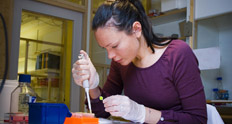
| Title | Microscopy: how to improve your imaging skills? Theory and practical tips from sample preparation to image analysis |
|---|---|
| Course number | 2820 |
| Programme | X- Används inte längre - Utvecklingsbiologi och cellulär signalering (DECS) |
| Language | English |
| Credits | 3.0 |
| Date | 2015-03-16 -- 2015-03-27 | Responsible KI department | Institutionen för biovetenskaper och näringslära |
| Specific entry requirements | Participants to this course must have used a microscope to acquire digital images of fluorescently labelled samples within the past 1 year. |
| Intended learning outcomes | At the end of the course, students will be able to: - Describe the difference between wide field and confocal microscopes as well as the different types of confocal microscopes and choose which system is most suited to which application - Evaluate fluorophores by matching their spectra with the microscope light source and filters, identify and avoid bleed-through and cross-excitation and pick the best combination of fluorophores for their own microscope - Explain objective specifications and limitations and describe the appropriate objective for their own application - Fix, mount and handle their sample in a way that is optimal for imaging as well as select an appropriate sample carrier for each application - Explain the theory behind resolution, pixel density, averaging, scan speed and describe which settings are best suited to their application - Explain which applications require a hardware autofocus, a spectral detector, a resonance scanner, two-photon microscopy or super resolution - Explain the advantages in using the automation of a microscope system to collect multidimensional data - Find their sample and the area of interest without bleaching it - Adjust the condenser for proper DIC imaging (Koehlering) - Run a simple image analysis and describe the imaging requirements for automated image analysis - Explain and critically comment on Material and Methods sections describing the acquisition of digital images and microscopy protocols |
| Contents of the course | The course will give students the theoretical and practical knowledge to improve the quality of their images, identify and avoid imaging artifacts and get started with automated image analysis. They will understand the difference between the different types of microscopy and which is required for which application. They will know what fluorescence is, how to understand fluorophore specifications, will have heard about the latest development in the fluorophore field and will be able to match fluorophore spectra and microscope filters to avoid bleed-through and cross-excitation. They will understand objective specifications and how to select the appropriate objective for their own application. They will learn about the challenges of imaging fluorescent volumes and the different techniques available. Participants will also understand how to set resolution, pixel density, averaging, scan speed, laser power, etc in the best way for their application. Additionally, they will hear many practical tricks about fixation, mounting and handling of their sample in a way that is optimal for imaging and they will learn about more advanced techniques like two-photon microscopy, super resolution, FLIM and spectral imaging. Lastly, they will get a chance to image their own sample, get feedback on what settings to use and improve their presentation and critical thinking skills. |
| Teaching and learning activities | Lectures, demonstrations, practical microscope lab sessions on State-of-the-Art confocals at the Live Cell Imaging microscopy facility, workshops and a minisymposium. |
| Compulsory elements | Attendance to all lectures and the labs is compulsory as well as passing the examination. In case of absence to a lab session, the student must present a literature work related to the missing activity. |
| Examination | The students will be assessed through a written examination (1h) with questions about the theoretical part of the course. Practical exercises will be marked during the workshops and hands-on. Students who fail will be given one extra chance for re-examination. |
| Literature and other teaching material | Reference literature: Handbook of Biological confocal microscopy, James Pawley, 2006 Recommended literature: Fluorescence microscopy ¿ avoiding the pitfalls, Claire M. Brown, Journal of Cell Science, 2007 Recent scientific papers and /or reviews presenting recent advances in the subject area will be provided at the start of the course. |
| Number of students | 12 - 16 |
| Selection of students | The selection is based on the usefulness of the course for the participant research projects and must be described in writing during the application. |
| More information | The course will be run on the latest Nikon confocals as well as a Zeiss LSM710-NLO (two photon) and an Andor spinning disk system. As far as possible, each participant should bring along their own sample as well as images they have acquired on their home microscope. Part of the homework is to image these samples again with the new knowledge in mind. Both theory and practice parts of the course will be held at the Live Cell Imaging unit, Karolinska Institutet, Huddinge mornings 9:00-12:00: lectures afternoons 13-17: days 1 and 9: workshop for all days 2-8: workshops and hands-on for half of the students or the other half (alternate days) day 10: a minisymposium with international speakers. Note that the date of the symposium may change depending on the availability of the speakers. It will be organized as close to the course as possible. |
| Additional course leader | |
| Latest course evaluation | Not available |
| Course responsible |
Sylvie Le Guyader Institutionen för biovetenskaper och näringslära Sylvie.Le.Guyader@ki.se |
| Contact person | - |